Blood Vessels Labeled Brain / Blood Vessel Model (with subtitles) - Ohio University ... - Microscopically, it is formed by the endothelium of the blood vessel.
Blood Vessels Labeled Brain / Blood Vessel Model (with subtitles) - Ohio University ... - Microscopically, it is formed by the endothelium of the blood vessel.. The blood vessels (and nerves) enter the brain through holes in the skull called foramina. Blood vessels innervate all tissues in vertebrates, enabling their survival by providing the necessary nutrients, oxygen, and hormonal signals. The two cell types ensure the integrity of the neural vasculature by maintaining the blood. Blood travels from the heart in arteries, which branch into smaller and smaller vessels, eventually becoming arterioles. If you use this material, please attach a link to the artwork, i would really love to see it :d.
Supplies the posterior brain, blood supply to the entire brain is ensured by anastomoses between the vessels. Blood vessels are vital for the body and play a key role in diabetes helping to transport glucose and insulin. In the cerebral medulla, the arteries and veins of the right side of the body are controlled from the left side of the brain; As well as providing new insights into the. About 2 years ago updated:
Migraines Linked With Abnormal Blood Vessel Structure In ... from i.huffpost.com Supplies the posterior brain, blood supply to the entire brain is ensured by anastomoses between the vessels. Blood vessels can be damaged by the effects of high blood glucose levels and this can in turn cause damage to organs, such as the heart and eyes, if significant blood vessel damage is sustained. In the article on the ventricles within the cns, we will discuss their structure and. If you use this material, please attach a link to the artwork, i would really love to see it :d. Cerebral arterial circle anterior communicating posterior cerebral a middle cerebral al reset zoom. Endothelial cells are labeled in red and pericytes in green. There is a right sided aca and a left sided aca. Another whole article within the blood vessels and csf section is dedicated to the cavernous sinus.
Cerebral arterial circle anterior communicating posterior cerebral a middle cerebral al reset zoom.
Comes off the subclavian a., ascends although the internal carotid a. In the cerebral medulla, the arteries and veins of the right side of the body are controlled from the left side of the brain; Only some of the vessels that exist in a real brain have been labeled. Over 1 year ago version: Blood vessels can be damaged by the effects of high blood glucose levels and this can in turn cause damage to organs, such as the heart and eyes, if significant blood vessel damage is sustained. The two cell types ensure the integrity of the neural vasculature by maintaining the blood. Endothelial cells are labeled in red and pericytes in green. Instead, transport is controlled mostly by the dilation of vessels. Blood vessels are intricate networks of hollow tubes that transport blood throughout the entire body so that it can deliver valuable nutrients to and remove waste from cells. Supplies the anterior brain and the vertebral a. The difference in the structural characteristics of arteries, capillaries and veins is attributable to their respective functions. Microscopically, it is formed by the endothelium of the blood vessel. Blood vessels are referred to collectively as the vascular system and, together with the heart, make up the circulatory system or cardiovascular system.
Blood is also supplied to the brain by the vertebral a. Label the blood vessels in the inferior view of the brain using the hints provided. Endothelial cells are labeled in red and pericytes in green. Cerebral arterial circle anterior communicating posterior cerebral a middle cerebral al reset zoom. • identification of blood vessels as arteries, capillaries or veins from the structure of their walls.
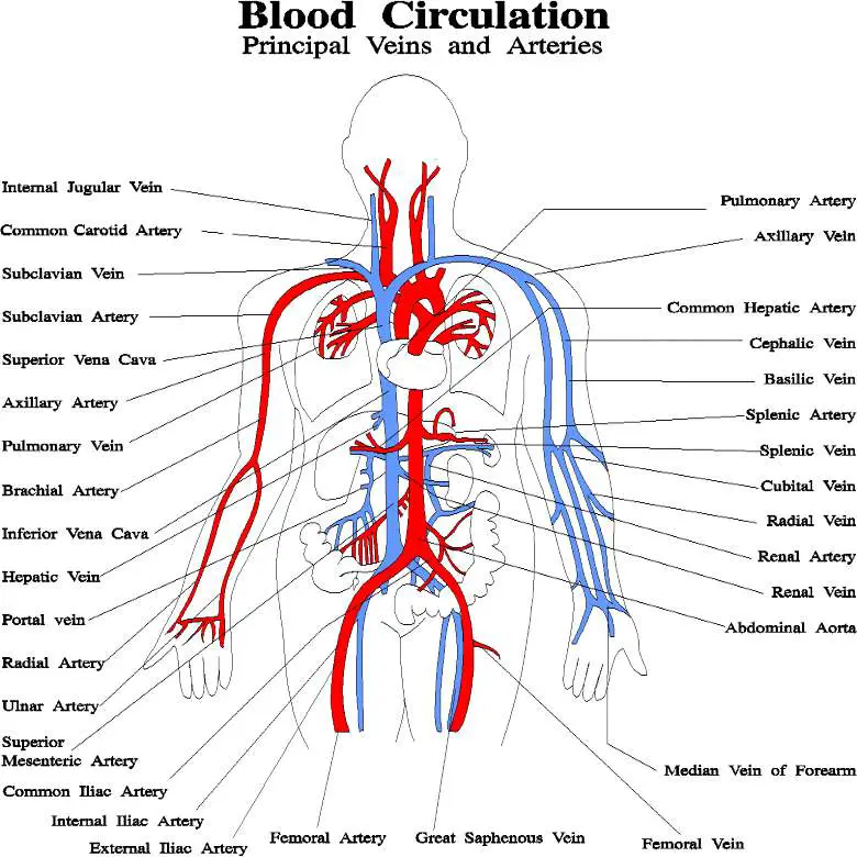
Blood vessels diagram from healthiack.com Blood travels from the heart in arteries, which branch into smaller and smaller vessels, eventually becoming arterioles. In the cerebral medulla, the arteries and veins of the right side of the body are controlled from the left side of the brain; Internal carotid artery (anterior circulation), vertebral artery (posterior circulation), and their hexagonal anastomotic network called blood brain barrier refers to the wall between the brain tissue and blood vessels. Only some of the vessels that exist in a real brain have been labeled. As well as providing new insights into the. Blood vessels are intricate networks of hollow tubes that transport blood throughout the entire body so that it can deliver valuable nutrients to and remove waste from cells. The difference in the structural characteristics of arteries, capillaries and veins is attributable to their respective functions. Instead, transport is controlled mostly by the dilation of vessels.
Researchers have discovered how cells of the blood vessels sense the metabolic condition of the brain and alter vascular function in response.
Internal carotid artery (anterior circulation), vertebral artery (posterior circulation), and their hexagonal anastomotic network called blood brain barrier refers to the wall between the brain tissue and blood vessels. The brain and its surrounding blood vessels exist in a close relationship. The blood vessels (and nerves) enter the brain through holes in the skull called foramina. Posterior communicating a internal carotid а. Blood vessels innervate all tissues in vertebrates, enabling their survival by providing the necessary nutrients, oxygen, and hormonal signals. Blood vessel with an erythrocyte (red blood cell, e) within its lumen, endothelial cells forming its tunica intima (inner layer), and pericytes forming its tunica researchers from the university of british columbia have discovered how blood vessels protect the brain during inflammation—a finding that. Equal to the intestinal muscles that move the food morsel along brain level: Supplies the anterior brain and the vertebral a. Microscopically, it is formed by the endothelium of the blood vessel. Label the veins of the anterior forearm and hand. Blood vessels are vital for the body and play a key role in diabetes helping to transport glucose and insulin. Researchers have discovered how cells of the blood vessels sense the metabolic condition of the brain and alter vascular function in response. Learn vocabulary, terms and more with flashcards, games and other study tools.
Blood vessels are vital for the body and play a key role in diabetes helping to transport glucose and insulin. In the article on the ventricles within the cns, we will discuss their structure and. Cerebral arterial circle anterior communicating posterior cerebral a middle cerebral al reset zoom. The carotid arteries and the vertebral arteries anterior cerebral artery (aca): The dense tight junctions between endothelial cells prevent paracellular transport through the.
Master blood vessels with quizzes and diagrams | Kenhub from thumbor.kenhub.com Blood is also supplied to the brain by the vertebral a. They also take waste and carbon dioxide away from the tissues. Label the veins of the anterior forearm and hand. There is a right sided aca and a left sided aca. The structure, distribution and labeling of the whole brain vascular system of different arteries and veins in 3d. • identification of blood vessels as arteries, capillaries or veins from the structure of their walls. Learn vocabulary, terms and more with flashcards, games and other study tools. Internal carotid artery (anterior circulation), vertebral artery (posterior circulation), and their hexagonal anastomotic network called blood brain barrier refers to the wall between the brain tissue and blood vessels.
Label the veins of the anterior forearm and hand.
Label the blood vessels in the inferior view of the brain using the hints provided. Posterior communicating a internal carotid а. The 500 ms patients, both adults and children, also underwent mri scans of the brain to measure iron deposits in surrounding areas of the brain. They also take waste and carbon dioxide away from the tissues. Label the blood vessels in the inferior view of the brain using the hints provided. Blood travels from the heart in arteries, which branch into smaller and smaller vessels, eventually becoming arterioles. Cerebral small vessel disease • not a single disease • group of diseases with different pathologies and different aetiologies • affecting the small arteries, arterioles, venules. Label the veins of the anterior forearm and hand. Supplies the anterior brain and the vertebral a. Red indicates arteries, and blue. This vessel supplies blood to the front part of your brain, knows as your frontal lobe. Blood vessels are vital for the body and play a key role in diabetes helping to transport glucose and insulin. The blood vessels (and nerves) enter the brain through holes in the skull called foramina.
Internal carotid artery (anterior circulation), vertebral artery (posterior circulation), and their hexagonal anastomotic network called blood brain barrier refers to the wall between the brain tissue and blood vessels blood vessels labeled. Instead, transport is controlled mostly by the dilation of vessels.


0 Komentar